
| Category | Cartilage Injury |
About Cartilage Injuries
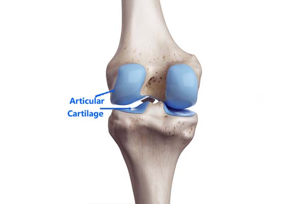
A normal joint has a layer of smooth white tissue covering the ends of the bones, known as Articular Cartilage. This layer of cartilage is vital for the proper functioning of the joint. Disruption of cartilage causes pain and restricts proper movement functioning of the joint.
Cartilage injuries can occur during injuries while playing in athletes, accidents, trauma, or can occur gradually over a period of months to years in patients with ligament/meniscal injuries or osteoarthritis. A special form of cartilage injury occurring in the patellofemoral joint, known as Chondromalacia Patellae affects the young population, more commonly females.
Symptoms
In the initial stages (Partial thickness injury) of cartilage injuries, the athlete complains of pain in the involved joint. The range of motion of the joint may also be limited due to pain. Joint swelling can also occur, sometimes related to physical activity.
The patient may also complain of clicks/crepitus or sounds from the joint while moving the involved joint. This is very common in Chondromalacia Patellae.
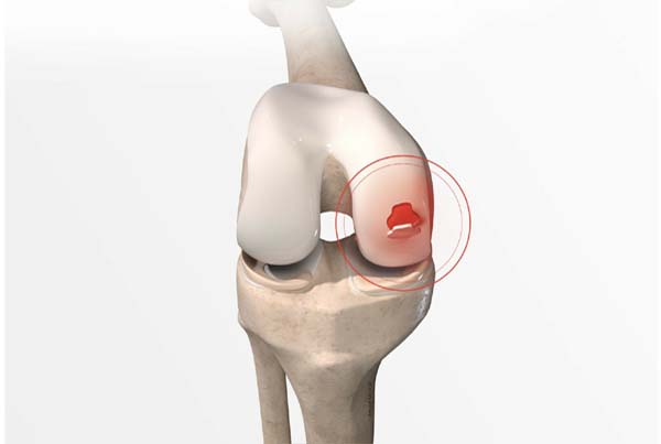
In full-thickness defects of cartilage, there might be a piece of cartilage that separates from the parent bone and act as a foreign body in the joint. It can then cause symptoms such as locking episodes, pain, and swelling of the joint.
Diagnosis
Clinical examination can raise a suspicion of a cartilage injury, but radiological investigations are necessary to localize and quantify the extent of the injury. X-rays are not of much use except in a few cases of severe/full-thickness injuries. MRI scan is the best modality to assess any cartilage damage currently. It helps to localize and stage the severity of the injury. Cartigram is an even more sensitive tool to pick up early cartilage changes.
Treatment
Partial-thickness cartilage injury is managed conservatively using medicines and rehabilitation. Some articular cartilage supplements like Diacerein, Glucosamine, Chondroitin, etc, can be used. Rehabilitation exercises focus on proper muscle conditioning and correcting any mechanical malalignment of the limb if present. Avoiding high-impact activities and postures that cause excessive stress on the cartilage is also recommended.
Complete thickness cartilage injury rarely improves with conservative treatment. These injuries require surgical treatment. There are various surgical techniques used to treat articular cartilage injuries.
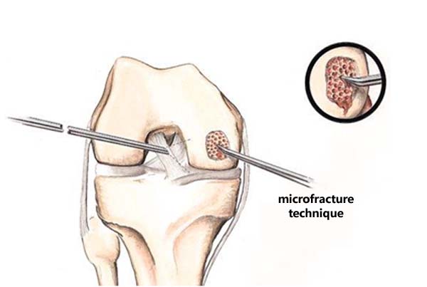
1. Microfracture
This arthroscopic technique is used to treat small-sized complete thickness cartilage defects. Multiple small-sized holes/ fractures are created in the underlying subchondral bone which allows the stem cells to help in cartilage healing.
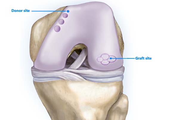
2. OATS ( Osteochondral Autologous Transfer System)
This arthroscopic technique involves the transfer of healthy cartilage with subchondral bone from the normal part of the joint to the affected area. The removed bone from the affected area is transferred back to the donor site. This technique is used to treat mild to moderate-sized lesions.
3. ACI
This technique is used to treat large-sized focal contained cartilage defects.
This is a 2 staged procedure. In first First stage, cartilage cells are harvested from the normal cartilage of the joint. This procedure is an arthroscopic procedure. These cartilage cells are then sent to a lab where they are grown in number in an artificial environment. This usually takes around 4 weeks.
In the second stage, the cultured cartilage cells embedded in special media are poured into the defect. This is an open procedure.
ACI is a relatively new technique with promising results, being practiced at a limited number of centers only.


